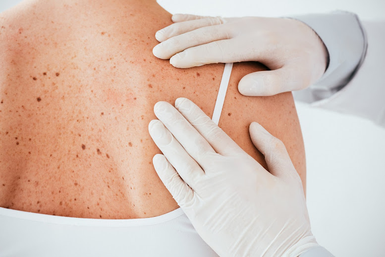
Dermoscopic Criteria for Melanoma
Melanoma is a serious form of skin cancer that arises from melanocytes, the pigment-producing cells of the skin. Early detection is crucial for effective treatment and improved patient outcomes. Dermoscopy Mole Evaluation in Dubai, a non-invasive imaging technique, plays a pivotal role in identifying melanoma through its specific dermoscopic criteria. This article discusses the ABCDE rule, additional dermoscopic features of melanoma, and the limitations of dermoscopy in melanoma detection.
The ABCDE Rule of Melanoma Detection
One of the most widely recognized methods for evaluating moles for potential melanoma is the ABCDE rule. This mnemonic aids both healthcare providers and patients in identifying concerning moles that require further investigation.
Asymmetry
- Definition: A mole is considered asymmetric when one half does not match the other in shape, color, or size.
- Implication: Asymmetry is a common indicator of malignancy. Benign moles typically exhibit symmetry, whereas melanomas often display irregular shapes.
Border Irregularity
- Definition: The borders of a mole are irregular when they are jagged, notched, or blurred.
- Implication: Melanomas often have poorly defined borders compared to benign moles, which usually have smooth, even edges.
Color Variation
- Definition: Color variation refers to the presence of multiple colors within a single mole, including shades of brown, black, red, white, or blue.
- Implication: Benign moles generally have a uniform color. In contrast, melanoma may show significant color variation, indicating a more aggressive lesion.
Diameter
- Definition: Diameter refers to the size of the mole. While melanomas can be smaller than 6mm, many exceed this size.
- Implication: Moles larger than 6mm, particularly those that exhibit other concerning features, warrant further evaluation.
Evolving Characteristics
- Definition: Evolving characteristics involve any changes in the mole's size, shape, color, or elevation over time.
- Implication: Changes in existing moles or the appearance of new moles in adults should be carefully monitored, as these can signal the development of melanoma.
Additional Dermoscopic Features of Melanoma
Beyond the ABCDE criteria, several other dermoscopic features can help in the identification of melanoma. Dermatologists are trained to recognize these characteristics, enhancing their diagnostic accuracy.
Dermoscopic Algorithms
Several dermoscopic algorithms have been developed to aid in the assessment of skin lesions. These algorithms use a combination of features to classify moles as benign or malignant. For example, the "Menzies Method" utilizes a checklist of features that includes asymmetry, border irregularity, and specific colors.
Importance of Vascular Patterns
The arrangement of blood vessels within a mole can also provide insights into its nature. Melanomas often exhibit atypical vascular structures, such as:
- Dot-like Vessels: Small, round, and irregularly shaped vessels can suggest malignancy.
- Branching Vessels: The presence of irregular branching vessels is often associated with more aggressive lesions.
Regression Structures
Regression structures refer to areas of depigmentation or lighter coloration within a mole. These areas may indicate that the body’s immune system is attacking the melanoma. While regression can occur in benign moles, it is more commonly associated with melanomas.
Limitations of Dermoscopy
While dermoscopy is a powerful tool for identifying melanoma, it is not without limitations. Recognizing these challenges is essential for accurate diagnosis.
False Positives and Negatives
- False Positives: Dermoscopy may incorrectly classify benign lesions as malignant, leading to unnecessary biopsies or treatments. This can occur in the presence of atypical nevi that exhibit features similar to melanoma.
- False Negatives: Conversely, some melanomas may not display the classic dermoscopic features, leading to missed diagnoses. Early-stage melanomas can sometimes present with subtle or atypical features that may be overlooked.
When to Consider a Biopsy
Dermoscopy is an invaluable preliminary step in mole evaluation, but when there is significant concern for melanoma, a biopsy may be necessary. Indications for biopsy include:
- A mole that exhibits multiple ABCDE features.
- A mole that has changed in appearance over time.
- A mole that shows concerning dermoscopic features, such as irregular vascular patterns or regression structures.
Conclusion
Dermoscopy is a crucial component in the early detection of melanoma, providing dermatologists with essential tools to identify concerning moles. By utilizing the ABCDE rule and recognizing additional dermoscopic features, healthcare professionals can make informed decisions regarding further evaluation and treatment. While dermoscopy has limitations, its role in the early detection of melanoma cannot be overstated, emphasizing the importance of regular skin checks and awareness of skin changes for better patient outcomes.


Comments
Post a Comment